surgical anatomy
MITRAL VALVE
Fibrous Skeleton of the Heart
- The overall structure and function of the heart depends on a widespread ‘honeycomb’ of connective tissue that courses throughout the heart, providing support to its cellular component.
- This is in turn supported by a more substantial network of dense connective tissue called the ‘fibrous skeleton of the heart'.
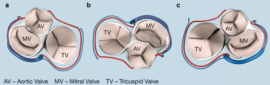
- It stabilizes the base of the ventricles and therefore provides a relatively inflexible but partially deformable scaffold for the annulus of the mitral, aortic and tricuspid valve.
- Serves as an electrical insulator between the atrial and ventricular component which is only interrupted at the AV node.
- Right Fibrous Trigone
Right fibrous trigone and Fila Coronaria
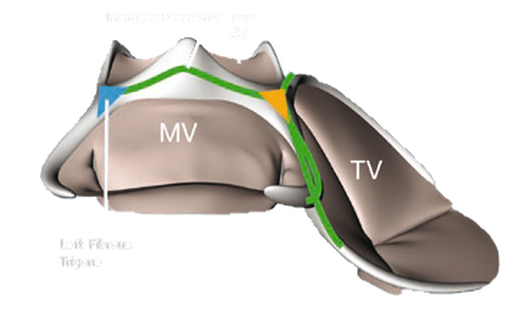
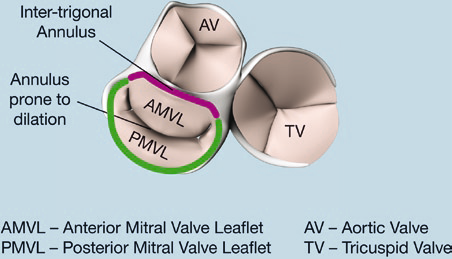
Superior, posteriorly directed Fila Coronaria forming the anterior mitral valve annulus
and “Inter-trigonal connective tissue”
and “Inter-trigonal connective tissue”
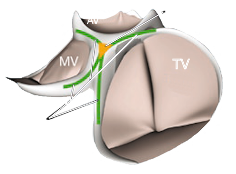
Left fibrous trigone in relation to the aortic valve
|
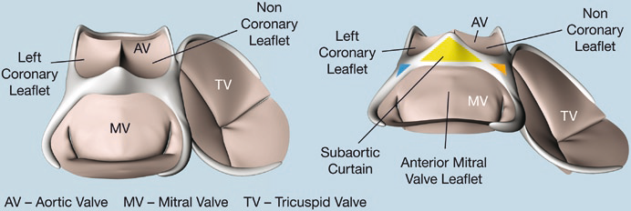
Aortic valve “Coronet”
|
Subaortic Curtain”
|
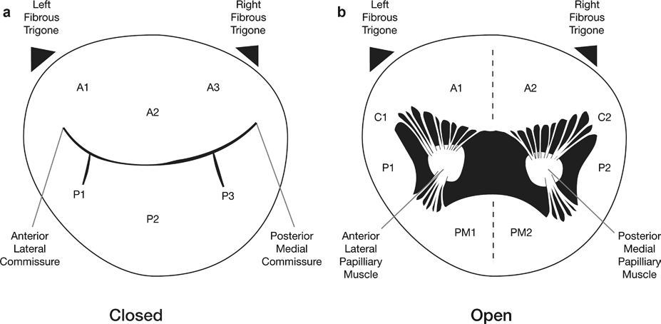
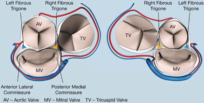
Relationship of trigones to the mitral valve commissures
|
Atrial aspect
|
Ventricular aspect
|