WALL SEGMENTS
The walls of the left ventricle are divided into three main segments with a further division into 16 sub-segments:
- Basal: subdivided into 4 sub-segments
- Medial: subdivided into 6 sub-segments
- Apical: subdivided into 6 sub-segments
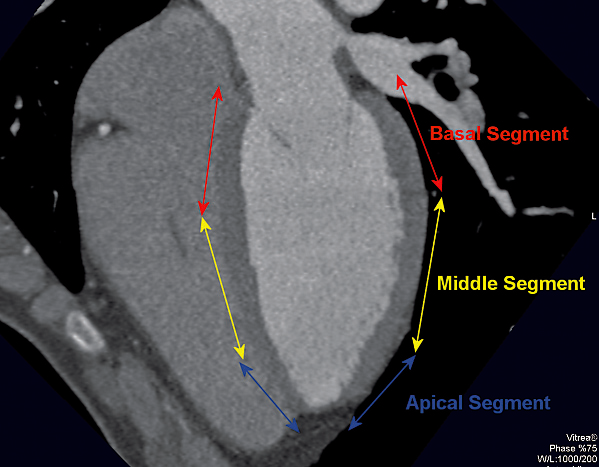
Four Chamber View
Subdivisions of the LV into three segments
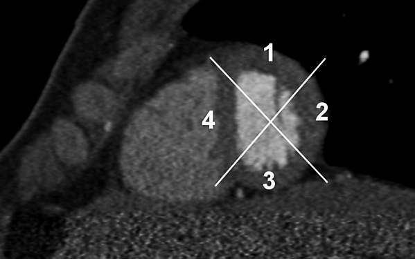
Short Axis View
Apical Segment divided into four sub-segments
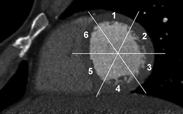
Short Axis View
Middle Segment divided into six sub-segments
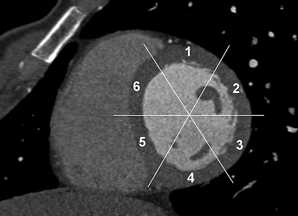
Short Axis View
Basal Segment divided into six sub-segments
ARTERIAL DISTRIBUTION PATTERN
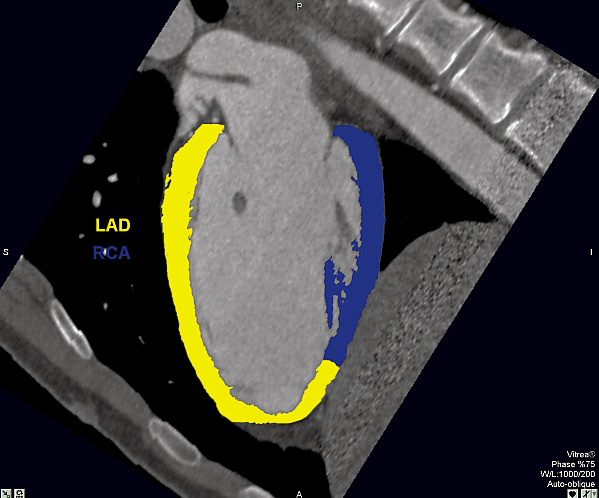
Two Chamber View
Most common distribution of the LAD (anterior wall) and RCA (posterior wall)
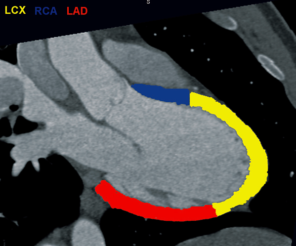
Three Chamber View
Most common distribution of the left circumflex, LAD and RCA

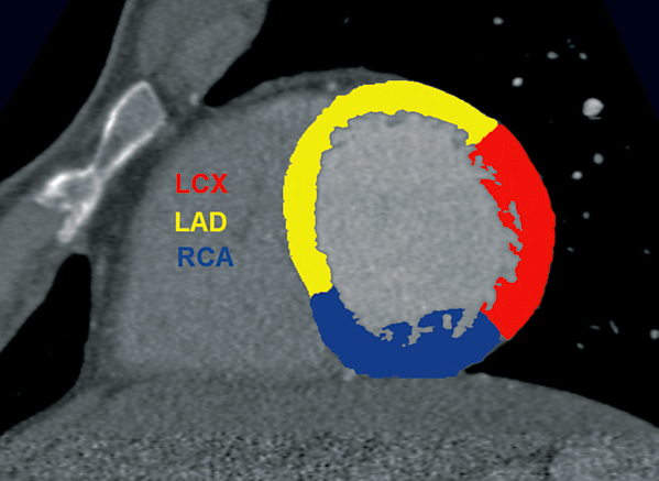
Four Chamber View
Most common distribution of the left circumflex, LAD and RCA
LCA - AP cranial (30°)
Most common distribution of the left circumflex (lateral wall),
LAD (anterior and septal wall) and RCA (inferior wall)
LAD (anterior and septal wall) and RCA (inferior wall)