CORONARY ANATOMY
anatomy of cardiac Veins
The cardiac veins may be divided into three groups:
- The largest system (Coronary Sinus and it tributaries), which collects a major amount of venous blood from the left ventricle and ends with the coronary sinus, opens into the right atrium.
- The second system (Anterior Right Ventricular veins) which gathers venous blood from the right two-thirds of the right ventricle and ends in the right atrium.
- The third is the system of the Thebessian veins, and comprises small veins that drain different portions of the right atrium and ventricle and empty directly and individually into these chambers. The internal openings, the foramina venarum minimarum, are best seen in the atrial septal wall
THE GREAT CARDIAC VEIN
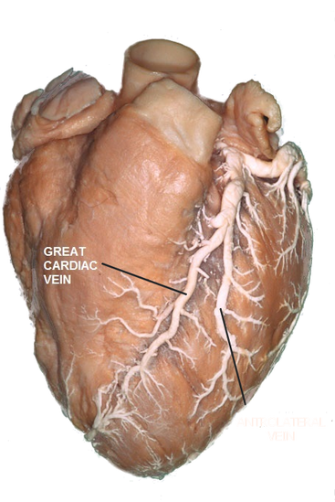
GREAT CARDIAC VEIN
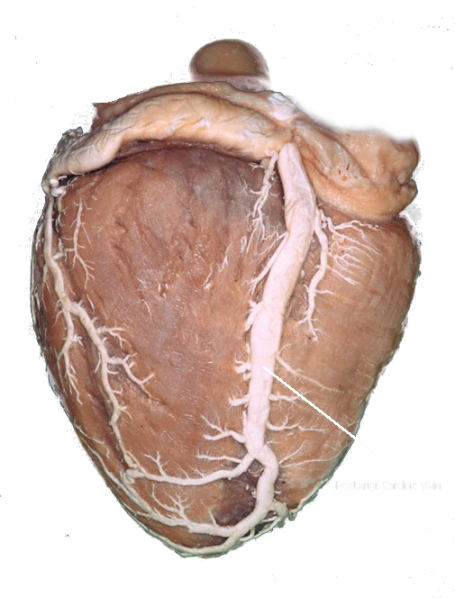
The anterior view presents the course of the great cardiac vein.
Starting in the lower third of the anterior interventricular sulcus and
runs toward the left coronary fossa
The Anterolateral vein is a large branch that drain the anterolateral surface
of the left ventricle and terminates by opening into the great cardiac vein
Starting in the lower third of the anterior interventricular sulcus and
runs toward the left coronary fossa
The Anterolateral vein is a large branch that drain the anterolateral surface
of the left ventricle and terminates by opening into the great cardiac vein
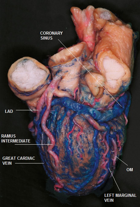
GREAT CARDIAC AND LEFT MARGINAL VEINS
Left lateral view of the Heart
On reaching the lateral wall of the pulmonary trunk, the great cardiac vein turns to the left and enters the left lateral extremity of the coronary sulcus.
Note that the left auricle covers the vein
Running beyond the left auricle, the vein reaches the margin of the left ventricle, and just beyond the obtuse margin the great cardiac vein opens into the coronary sinus
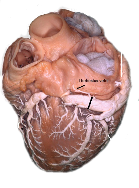
Left lateral view of the transition between the great cardiac vein and the coronary sinus
Solid black line is positioned between coronary sinus and great cardiac vein
Solid black line is positioned between coronary sinus and great cardiac vein
The transition between the coronary sinus and the great cardiac vein is marked by two morphological structures:
1. The valve of Thebesius (also known as the valve of Vieussens)
2. The Thebesius vein (The oblique vein of the left atrium) (also known as the vein of Marshall)
A relatively large marginal vein is seen; this vessel collects the venous drainage from the lateral left ventricular wall, running at the obtuse margin and finally opening into the coronary sinus
1. The valve of Thebesius (also known as the valve of Vieussens)
2. The Thebesius vein (The oblique vein of the left atrium) (also known as the vein of Marshall)
- The vein runs along the anterior surface of the left atrium toward the coronary sinus
- This small vein is present in 95% of cases
- It has a diameter of up to 1 mm
- It passes between the left appendage and the left inferior pulmonary vein toward the roof of the left atrium
- Embryologically: the vein develops from the left superior cardinal vein, and in some cases persists in adults as the left superior vena cava which is commonly associated with congenital defects such as tetralogy of Fallot (incidence of 20%) and Eisenmenger’s syndrome (incidence of 8.3%), and is frequently an isolated malformation that receives the hemiazygos vein
A relatively large marginal vein is seen; this vessel collects the venous drainage from the lateral left ventricular wall, running at the obtuse margin and finally opening into the coronary sinus
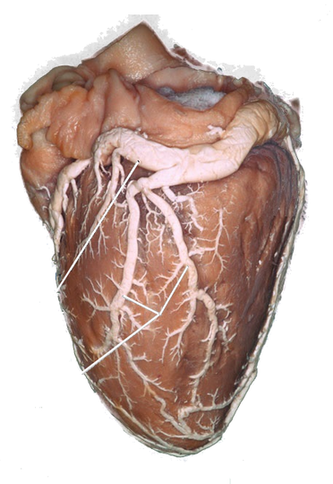
Transition between the great cardiac vein and the coronary sinus in the left coronary fossa
- After passing the lateral wall of the pulmonary root, the great cardiac vein runs straight toward the left coronary fossa.
- On reaching the anterior wall of the left atrium, the great cardiac vein turns at a right angle and runs further into the coronary sinus in front of the anterior atrial wall, close to the mitral wall.
- To left of the left atrium, a string-like branch opens into the great cardiac vein, collecting the venous drainage from the area of the coronary fossa. This small venous branch wraps around the left coronary sinus, an area corresponding to the superior septal area. This branch probably collects the venous drainage from the superior part of the interventricular septum.
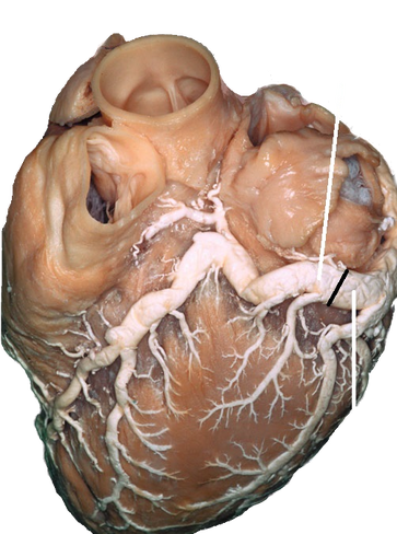
THE MIDDLE (POSTERIOR) CARDIAC VEIN
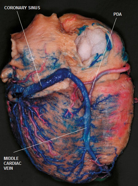
Posterior cardiac vein, posterior view
- The posterior cardiac vein (posterior interventricular vein or the middle cardiac vein) commences at the apex of the heart and ascends along the posterior interventricular groove parallel to the posterior descending artery (PDA) toward the coronary sinus up to the crux. It opens directly into the coronary sinus or into the right atrium
- The posterolateral and the obtuse marginal veins both open into the coronary sinus.
- The posterolateral vein is placed at the diaphragmal surface of the heart.
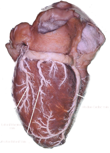
Lateral view of the cardiac venous system
- The marginal vein is presented as a greater branch running at the obtuse margin, collecting the venous drainage from the anterior and diaphragmal surfaces of the heart.
- The small posterolateral veins collect the venous blood from the diaphragmal surface of the left ventricle and are positioned between the marginal vein and the posterior cardiac vein
THE SMALL ANTERIOR CARDIAC VEINS

The anterior small cardiac veins
Small Anterior Cardiac Veins
- Drain the anterior right ventricular wall
- Run in the pericardial fat tissue
- Bridge the RCA from the superior side
- Open directly into the right atrium
- Runs parallel to the right marginal artery and over the right diaphragm.
- Collects the blood from the anterolateral wall of the right ventricle
- Opens directly into the right atrium
THE CORONARY SINUS
Coronary sinus with cardiac veins on the diaphragmatic surface of the heart
Posterior view of the coronary sinus
- The coronary sinus predominantly drains the left ventricle and receives approximately 85% of coronary venous blood.
- It lies within the posterior atrioventricular groove and empties into the right atrium at the lateral border of the triangle of Koch
- The orifice of the coronary sinus is guarded by the crescent-shaped thebesian valve.
- The named tributaries of the coronary sinus include the anterior interventricular vein, which courses parallel to the left anterior descending coronary artery. Adjacent to the bifurcation of the left main stem, the anterior interventricular vein courses leftward in the atrioventricular groove, where it is referred to as the great cardiac vein
- The ostium of the coronary sinus is observed just to the right of the posterior interventricular groove
- This corresponds to the left margin of the superior posterior process of the left ventricle
- The position of the coronary sinus ostium also influences the course of the posterior coronary vein. (a right-sided ostium results in a right-sided posterior cardiac vein, and vice versa, relative to the posterior interventricular sulcus)
- In its course over the posterior groove, the vein covers the posterior descending branch (PDA)
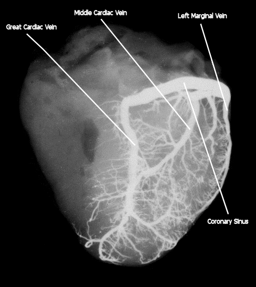
Angiogram of the heart veins
- The coronary sinus collects the entire venous drainage from the left ventricle via the great and middle cardiac veins
- In addition, the left marginal and posterolateral veins contribute to the left ventricular venous drainage
- The small cardiac vein is present as a small vein that opens into the terminal part of the coronary sinus
- Clinical Importance:
muscles as the veins of the right ventricle do not communicate with the coronary sinus, and so the
cardioplegia can not reach the right half of the heart
- This may be related to the variable venous anatomy of the heart; because the anterior region of
the right ventricle is not drained by the coronary sinus, and it is not uncommon for the heart to
have several coronary sinus anomalies, there may be a heterogeneous distribution of cardioplegic
solutions, thus limiting myocardial protection. As a consequence, a technique for simultaneously
delivering cardioplegia both antegradely and retrogradely has been developed
RELATIONSHIP BETWEEN THE CORONARY ARTERIES AND VEINS
The Anterior Great Cardiac Vein and the LAD:
Starting at the apex:
Starting at the apex:
- The anterior great vein runs on the left side of the LAD toward the left coronary Fossa.
- It should be noted that the small venous branches providing the drainage of the right and left ventricles may cross the LAD anteriorly before joining the great vein which can be a source of annoying bleeding during surgical exposure of the LAD
- It has been postulated that in most cases the great cardiac vein runs on the right side of the artery
- In some cases there may even be two venous branches commencing on both sides of the LAD.
- Rarely, the course of the anterior great cardiac vein lies over the LAD which does affect the surgical exposure the artery, espciially when the epicardium is covered by a large amount of fat tissue, in which case the surgeon may mistake the vein for the artery.
- This misinterpretation may also result from an intramural position of the LAD
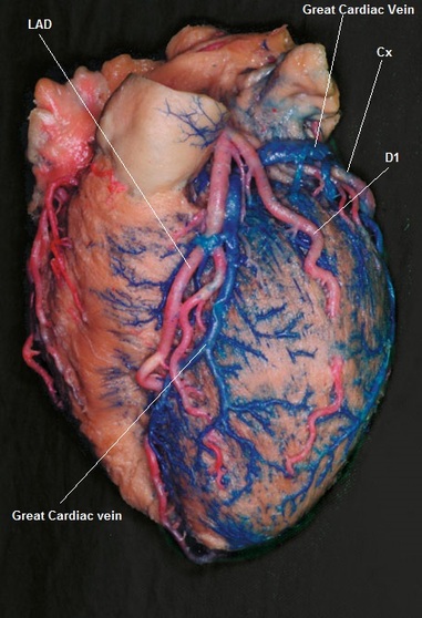
The relation between the Great Cardiac Vein and the LAD
The Anterior Great Cardiac Vein and the Circumflex artery (Cx):
- The great cardiac vein runs on the left side of the retro-pulmonary part of the LAD
- At the termination of the anterior sulcus, the vein turns to the left toward the coronary sulcus.
- In most cases, the vein crosses the Cx superiorly in the left coronary fossa.
- In the initial part of the coronary sinus, the vein has a very close relationship with both the left atrium and the mitral valve.
- The Cx is positioned under the great cardiac vein, on edge of the left ventricular ostium
- In this morphological situation, the artery cannot be identified at this level.
- However, on reaching the area of the obtuse margin, the vein runs smoothly from the level of the ventricular wall to the wall of the left atrium.
- As a consequence, the Cx becomes superficial and may be easily identified just inferior to the coronary sinus.
- Clinically; the great vein reaches the ostium of the left ventricle at the level of the left fibrous trigone, thus the vein is prone to injury during mitral or aortic valve surgery.
- In some cases the vein may interdigitate with both the LAD and the Cx. Making the intra-operative identification of the loop formation in this area obviously discourages creating any graft anastomosis at the initial part of the LAD and Cx.
The relation between the Great Cardiac Vein and the Circumflex artery
- Due to superior dislocation of the great cardiac veins, the coronary sinus finally runs superior to the attachment of the left atrium.The Cx is positioned just inferior to the sinus, in the coronary groove.
- The marginal and posterolateral veins run superior to the artery.
- The marginal and posterolateral veins are not always so prominent.
- Furthermore, the veins on the posterior wall of the left ventricle are not ordinarily adjacent to the arteries and cannot be used as a guide for the identification of the latter.
- However, at the obtuse margin the marginal vein is always in contact with the artery.
Anatomical Location of Coronary Arteries and Veins
|
Coronary Vein
|
Anatomical Location
|
Coronary Artery
|