NORMAL CT CORONARY ANATOMY

Three Dimensional CT Image
3D Image showing RCA and LAD
Three Dimensional CT Image
3D CT image of the ascending aorta and the coronary artery tree, with transparent other cardiac structures

Maximum intensity projection (MIP)
Maximum intensity projection (MIP) showing RCA, LAD,a diagonal of the LAD and the circumflex artery (CX)

Course of LAD
LAD coursing from the anterior wall of the left ventricle to the apex

Axial CT slide
Axial CT slide showing the bifurcation of the LAD and the CX artery
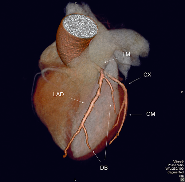
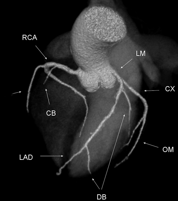
Left Coronary Artery System
Left Coronary Artery System
LM, left main coronary artery; CX, circumflex artery;
OM, obtuse marginal branch; DB, diagonal branches;
LAD, left anterior descending artery
LM, left main coronary artery; CX, circumflex artery;
OM, obtuse marginal branch; DB, diagonal branches;
LAD, left anterior descending artery
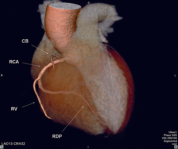
Right Coronary Artery System
Right Coronary Artery System
CB, conus branch;
RVB, right ventricular branch;
RDP, right descending posterior branch
CB, conus branch;
RVB, right ventricular branch;
RDP, right descending posterior branch
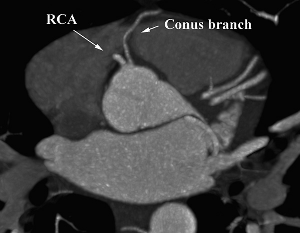
Maximum Intensity Projection (MIP)
Maximum Intensity Projection (MIP) of a cardiac CT
showing the separate origin of the RCA and conus branch
showing the separate origin of the RCA and conus branch